
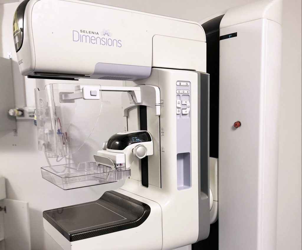
Metrological evaluation of these advanced mammography systems requires rigorous characterization of the clinical X-ray beams used. To achieve this, our Henri Becquerel National Laboratory (LNHB-MD) replicated the beam configurations used at the Georges Pompidou European Hospital (HEGP).
Through sophisticated analysis and correction procedures that address escape effects caused by fluorescence and pile-up effects caused by high counting rates, the team reproduced clinical spectra with exceptional precision. These beams now enable the LNHB-MD to calibrate clinical radiameters, ensuring accurate monitoring of patient exposure levels during diagnostic exams.
This makes it possible to balance diagnostic image quality with the varying radiation dose requirements of individual breast anatomy.
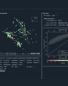
This research is part of the national metrology program, a collaboration with HEGP and a doctoral thesis aimed at automating the measurement of mean glandular dose, the indicator for optimizing patient exposure. This tool will enhance dose traceability during mammograms and improve the safety of tomosynthesis in breast cancer screening.
million women were diagnosed with breast cancer in 2022
deaths globally in 2022. Source: WHO website
Tomography invites us to rethink the metrological traceability chain, from defining the dosimetric quantity to the measurement methods.
By improving our knowledge of the doses delivered to patients, this collaboration helps make using breast tomosynthesis for breast cancer screening safer.